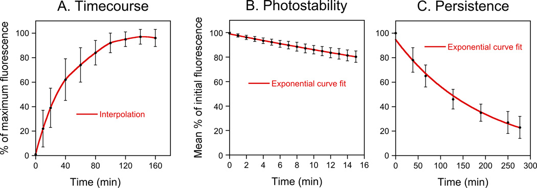
Fig. 5. Characterization of FluoroMyelin™ Red staining.
A. FluoroMyelin™ Red staining time-course. Data from 28 myelin sheaths in 5 dishes that were stained after 31–41 days in culture. B. FluoroMyelin™ Red photostability. The culture was stained for 2 hours and then the excess dye was rinsed off with fresh observation medium and each field of view was imaged continuously for 15 minutes with the light from the mercury arc lamp attenuated 12-fold using neutral density filters. Data from 46 myelin sheaths from 6 different fields of view in one dish that was stained after 67 days in culture. C. Persistence of FluoroMyelin™ Red staining after wash-out. The culture was stained for 2 hours and then the excess dye was rinsed off with fresh observation medium and selected myelin sheaths were imaged at 30–60 minute intervals for 5 hours. Data from 14 different myelin sheaths in one dish that was stained after 45 days in culture.

