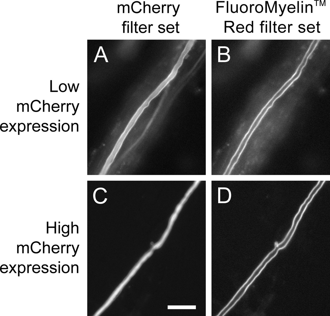
Fig. 8. Separation of FluoroMyelin™ Red and mCherry fluorescence using a custom FluoroMyelin™ Red filter cube.
A myelinating culture was transfected with mCherry after 40–47 days in culture. Four days later, the culture was exposed to FluoroMyelin™ Red (1:300 dilution) in observation medium for 2 hours, and then rinsed and imaged in observation medium with a 40× objective magnification. A,B. A myelinated axon expressing relatively low levels of mCherry. C,D. A myelinated axon expressing relatively high levels of mCherry. Both mCherry and FluoroMyelin™ Red fluorescence are visible with the mCherry filter set (A,C) because both dyes excite in the green and emit in the red, but only the FluoroMyelin™ Red fluorescence is visible using the custom FluoroMyelin™ Red filter set (B,D) because mCherry does not excite in the blue, whereas FluoroMyelin™ Red does. When the mCherry expression is relatively low, the myelin sheath appears on the mCherry channel as a bright border along either side of a faint axon (A), whereas when the mCherry expression is relatively high the myelin sheath appears on the mCherry channel as a faint halo along either side of a bright axon (C). Scale bar= 10 µm.

