
Figure 2.
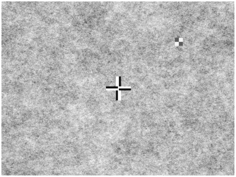
Patient’s view during testing consisting of a patterned background (1/f 0.75 spatial noise), a bipolar fixation cross, and a peripheral bipolar checkerboard target (shown here at high, 95% contrast). Only the central portion of the screen is shown.