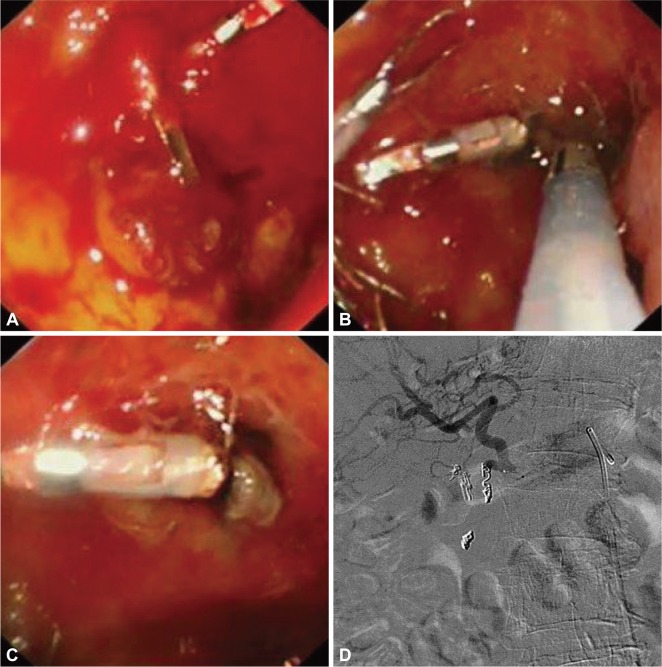
Fig. 4.
A large bulbar ulcer that failed hemoclipping (A) was treated by thermo-coagulation using a 3.2 mm heater probe (B, C). The gastroduodenal artery was then coiled during angiography (D). The picture depicts a microcatheter in the common hepatic artery and a larger Simmon's catheter in the celiac artery. The hemoclips provide landmark to the site for empirical coiling. Coils are first dropped distal to the bleeding point. Gelfoams are then used to block collateral branches. Further coils are then added to the proximal portion of the gastro-duodenal artery.

