
Figure 6.
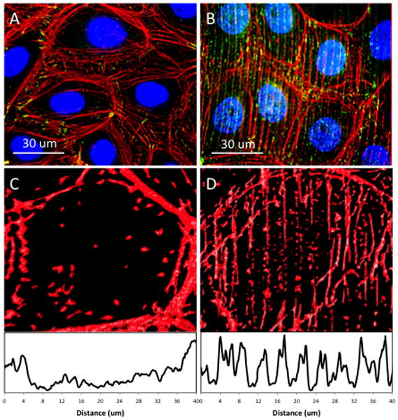
Fluorescent microscopy images of the cytoskeletal localization (red – actin, green – vinculin) upon both (A) flat and (B) patterned silk film surfaces. Representative images of cell actin localization upon (C) flat and (D) lined silk film surfaces with respective surface plot profiles shown below demonstrating actin fibril localization towards the periphery on flat silk film surface, and fibril alignment along the patterned lined silk topography as indicated by the periodical intensity peaks corresponding to feature presence.