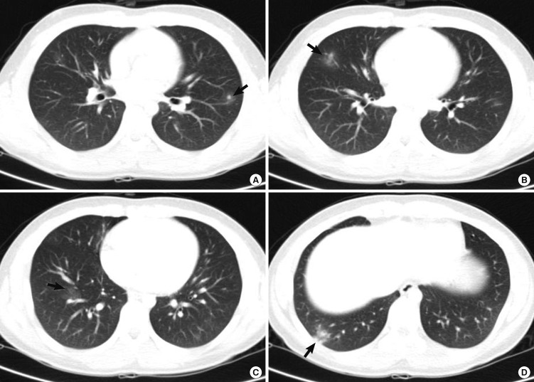
Fig. 1.
Computed tomography (CT) scan findings of the chest. Multiple nodules with ground glass opacity halo in the left upper lung field (A) and ill defined patches of nodular ground glass opacity are shown in the right middle (B) and the right lower lung field (C, D), which involve mainly the peripheral regions of the both lungs (arrows).

