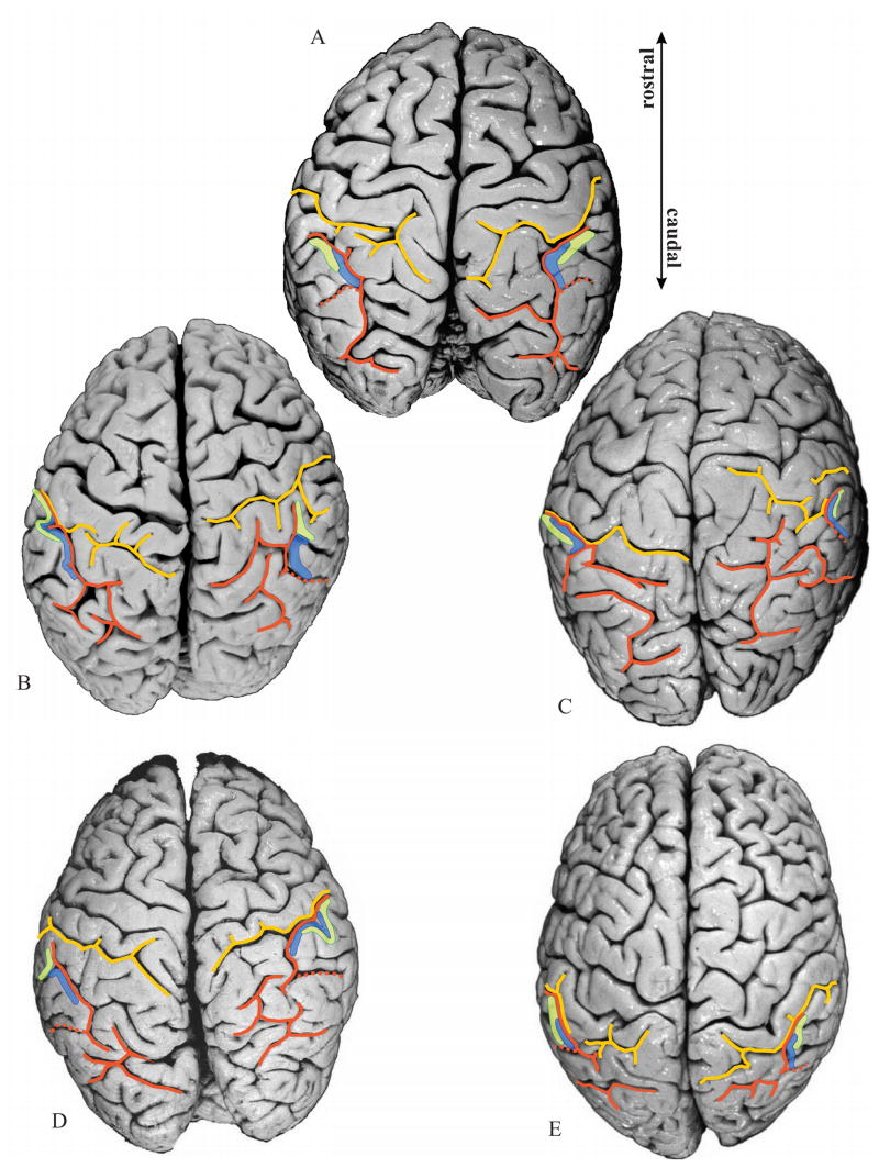
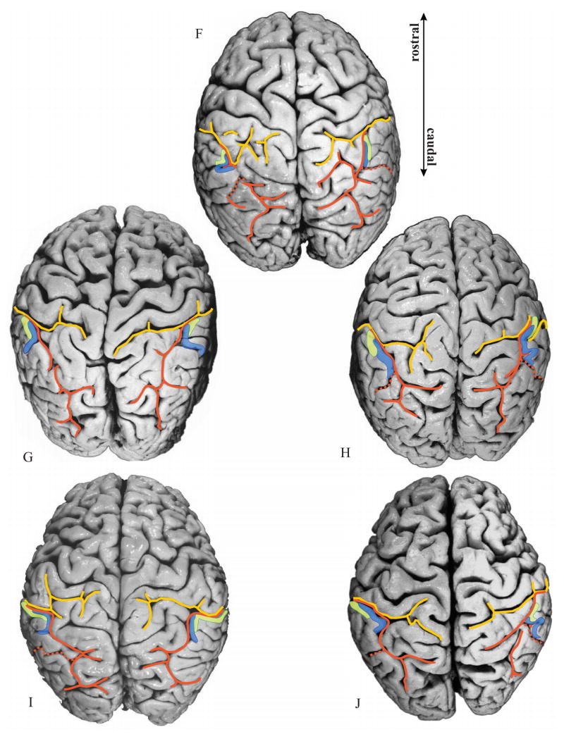
Figure 7.
Delineated areas hIP1 (blue) and hIP2 (green) are projected onto photographs of the dorsal surface of the 10 delineated postmortem brains. Note the considerable intersubject variability in the pattern of the IPS (orange) and PCS (yellow), as well as the variability in location and extent of both areas. Orange dotted line: IMPS, the side branch of the IPS, which divides the supramarginal gyrus from the angular gyrus. Note that in the caudal direction hIP1 does not extend beyond this side branch.


