
Fig. 3.
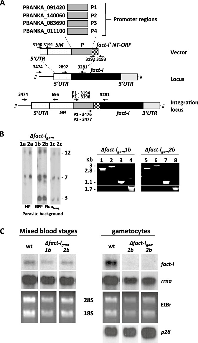
Development of a ‘promoter-swap’ technology for replacing the fact-l promoter with an ‘asexual-specific’ promoter. A. Schematic representation of the promoter-swap vector and the promoter regions of four selected genes, P1–P4. This vector integrates into the fact-l locus through a double-cross-over integration event resulting in replacement of part of the fact-l promoter region with the tgdhfr-ts selectable marker (SM) and one of the P1-P4 promoters. The location of primers for diagnostic PCRs (see B) for correct integration is shown. Fact-l' NT-ORF represents the N-terminal encoding region of fact-l used in the vector to ensure correct integration. B. Left panel: Southern analysis of separated chromosomes of six independent Δfact-lgam mutants showing correct integration of the promoter-swap vectors containing promoter P1 (pL1312; 1a–c) or promoter P2 (pL1313; 2a–c). Hybridization with the 3′-dhfr/ts probe recognizes the construct integrated into the fact-l locus on chromosome 12, the endogenous dhft/ts gene on chromosome 7 and the transgenes of the parent parasite lines wtGFP and wt-Fluofrmg integrated into chromosome 3 (GFP in 1b, 2b and GFP/RFP in 1c, 2c). Upper panels: Diagnostic PCR analysis showing correct integration of vectors containing promoter P1 and P2 in Δfact-lgam 1b and Δfact-lgam 2b respectively (see A and Table S1 for primer location and sequence). Lanes 1, 5: primers 3474/695; lane 2, 3: promoter P1, primers 3194/3281 and primers 3476/3281; promoter P2, lane 6: primers 3196/3281; lane 7: primers 3477/3281. Amplification of the wild-type FACT promoter region is negative in Δfact-lgam 1b and Δfact-lgam 2b lanes 4, 8 respectively using primers 2892/3281. Lower panels: Amplifications using the same primer pairs indicated for the upper panels with wt PB ANKA DNA as template. C. Northern analyses of fact-l transcription in wild type and two Δfact-lgam mutants, showing the presence of the two Δfact-lgam mutants in mixed blood stages but the absence of fact-l transcripts in purified Δfact-lgam gametocytes; hybridization of p28 is included to indicate similar amount of gametocyte RNA loading.