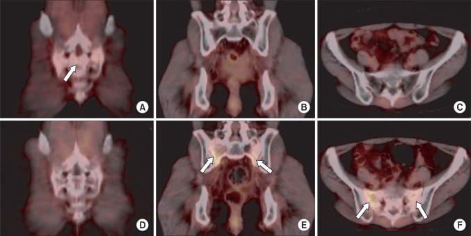
Fig. 3.
A 48-year-old woman (patient 3) who received definitive chemoradiotherapy for cervical cancer developed a hip pain at 12 months after radiotherapy. Serial positron emission tomography/computed tomography (PET/CT) scans at 12 months after radiotherapy (A-C) and 21 months after radiotherapy (D-F). A transverse linear mild flourodeoxyglucose (FDG) uptake (SUVmax, 1.7) in the sacrum (arrow) was shown on coronal fusion image at 12 months after radiotherapy (A). Follow-up PET/CT at 21 months after radiotherapy showed normalized transverse linear uptake (D) which was seen on previous PET/CT. A new vertical linear mild FDG uptake parallel to bilateral sacral alae (arrow) was shown on 21-month follow-up PET/CT (E,F), in which previous PET/CT scan showed normal finding (B,C). SUVmax, maximum standardized uptake value.

