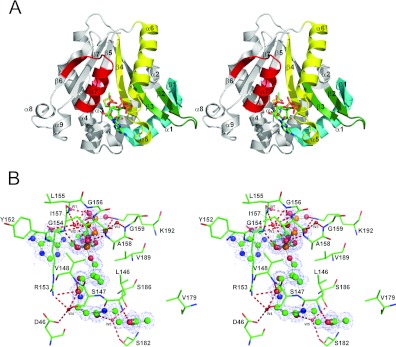
Figure 1. The crystal structure of the Dat21–230–AcCoA complex and interactions between Dat21–230 and AcCoA.
(A) A stereo ribbon diagram of the Dat21–230–AcCoA complex. Dat contains nine α-helices and seven β-strands. The conserved GNAT motifs C, D, A and B are coloured cyan, green, yellow and red respectively. AcCoA is shown as a stick model and is situated in its binding cavity. (B) A stereo 2Fo−Fc electron density map for AcCoA contoured at 1 σ within the stick model of the AcCoA-binding site. Hydrogen bonds are shown as broken lines. A certain number of the Dat21–230–AcCoA interactions are mediated by hydrogen bonding with water molecules (W1–W5, small red balls).

