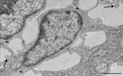
Figure 6. Transmission electron micrograph of embryonic E21 rat tail tendon.
The image shows fibricarriers (collagen fibrils within the main body of the cells) (short arrows) and fibripositors (collagen fibrils located within cellular projections) (long arrows), in transverse view. Fibripositors and fibricarriers are characteristic features of embryonic tendon. Scale bar, 1 μm.

