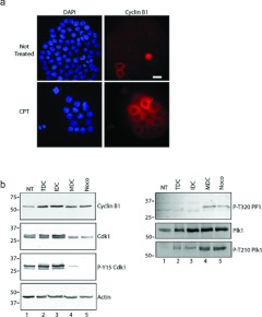
Figure 3. MDCs express cyclin B1 and the active form of Cdk1.
(a) Cells were grown on coverslips and either not treated (upper row) or treated with CPT for 48 h (lower row), stained with DAPI (blue) and with anti-(cyclin B1) antibodies (red). Cells were analysed by immunofluorescence microscopy. Scale bar, 50 μm. (b) Extracts were prepared from cells either not treated (NT) or treated with CPT. CPT-treated cells were analysed further as total cultures (TDC) or cultures fractionated by mechanical shake-off into adherent interphase cells (IDC) or rounded cells (MDC). Extracts were also prepared from cells treated with nocodazole (Noco). Samples were processed by Western blotting with antibodies against cyclin B1, Cdk1, phospho-Tyr15 Cdk1 (P-Y15 Cdk1), phospho-Thr320 PP1a (P-T320 PP1), Plk1, phospho-Thr210 Plk1 (P-T210 Plk1) or actin. Molecular masses are indicated in kDa.

