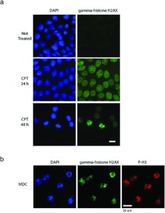
Figure 6. MDCs are in mitosis with damaged DNA.
(a) Cells were either not treated or treated with CPT and stained with DAPI (left-hand panels) and γH2AX antibodies (right-hand panels). Cells were observed after 24 and 48 h of treatment by immunofluorescence microscopy. Scale bar, 10 μm. (b) MDCs were collected, attached to coverslips and stained with DAPI (left) and antibodies against γH2AX (centre) or phospho-Ser10 histone H3 (right, P-H3) and observed by confocal microscopy. Scale bar, 20 μm.

