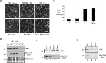
Figure 7. Chk1 is dephosphorylated and not degraded in cells in mitosis with damaged DNA.
Cells were either not treated or treated with MG132 for 6 h or with CPT for 48 h. At 48 h, MDCs were removed by mechanical shake-off from a total culture and the remaining IDCs were either re-incubated with CPT for 6 h (IDC 54 h) or incubated with CPT and MG132 for 6 h (IDC + MG132 54 h). Scale bar, 100 μm. (b) The number of MDCs collected by mechanical shake-off was counted at 48 h under each treatment condition and presented as percentages relative to non-treated cells (NT). (c) Cells were either not treated (NT) or treated with increasing concentrations of CPT (half-log; 0.03–3 μM). Extracts were prepared and analysed by Western blotting with antibodies against either Chk1 or phospho-Ser345 Chk1 or by Coomassie Blue staining. Molecular masses are indicated in kDa. (d) Cells were treated with CPT for 48 h (TDC) and separated into IDC and MDC populations. Mitotic cells without damaged DNA were prepared from nocodazole-treated culture (Noco). Samples were processed by Western blotting with antibodies against either Chk1 or phospho-Ser345 Chk1. Molecular masses are indicated in kDa. (e) Cells were treated with CPT for 48 h (TDC) and separated into IDC and MDC populations (MDC1). The IDC cells were re-cultivated for 2 h and new mitotic cells were collected (MDC2). Samples were processed by Western blotting with antibodies against either Chk1 or phospho-Ser345 Chk1. Molecular masses are indicated in kDa. p, phospho-

