Figure 1.
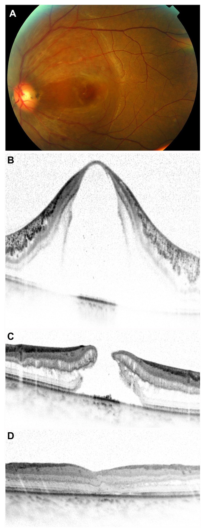
Case 3. Preoperative fundus photograph showing a temporally located optic disc pit with associated maculopathy (A). Spectral-domain optical coherent tomography (Spectralis, Heidelberg Engineering, Heidelberg, Germany) images through the macula obtained before (B), 2 months after (C), and 3 months after (D) first vitrectomy.

