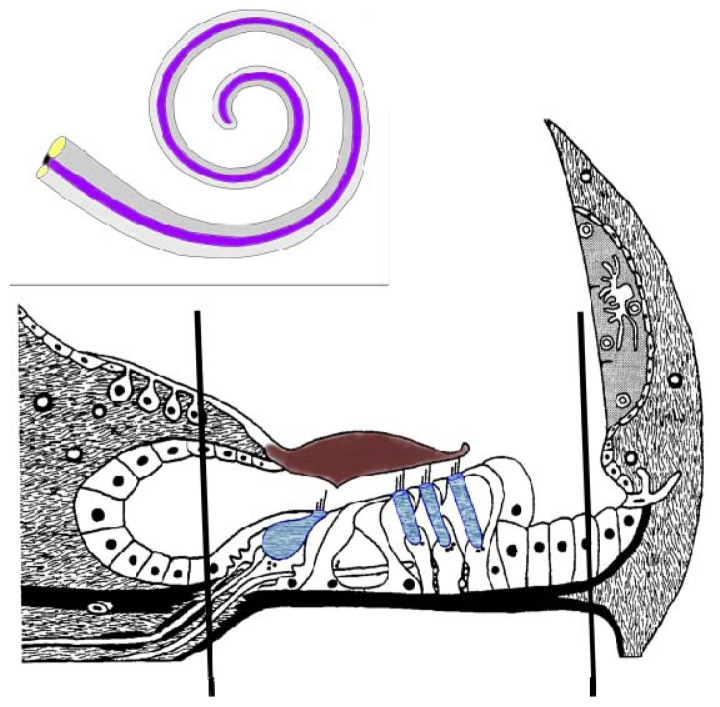
Figure 1.
Schematic representation of the cochlear membranous labyrinth and the organ of Corti. A cross section of the cochlea is represented with the hair cells (blue) being depicted with the single inner hair cell and three outer inner hairs. The black lines represent the approximate region isolated by dissection for this proteomic investigation. The framed insert represents the membranous labyrinth of the cochlea with the anterior scala vestibuli (gray), the scala media (cochlear duct) (purple) that contains the organ of Corti and the scala tympani (light gray).

