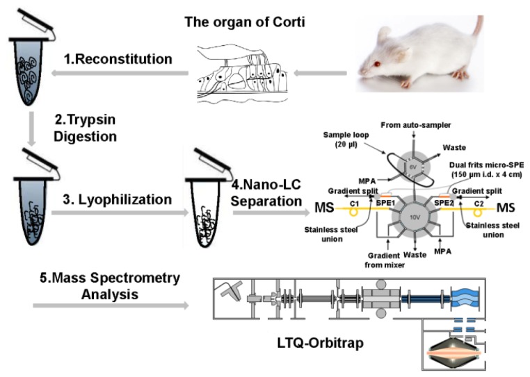
Figure 2.
Schematic diagram of proteomic analysis of the mouse organ of Corti (OC) sample. (1) The OCs were reconstituted in 100 μL lysis buffer (100 mM ammonium bicarbonate, pH 8.4); (2) The lysates were reduced by 5 mM DL-Dithiothreitol (DTT) and digested by trypsin overnight; (3) The digests were desalted and dried in a vacuum centrifuge immediately after digestion; (4) Dried peptides were subjected to the in-house assembled reverse phase metal-free multiple-column nanoLC system coupled with (5) LTQ-Orbitrap XL mass spectrometer for MS analysis.

