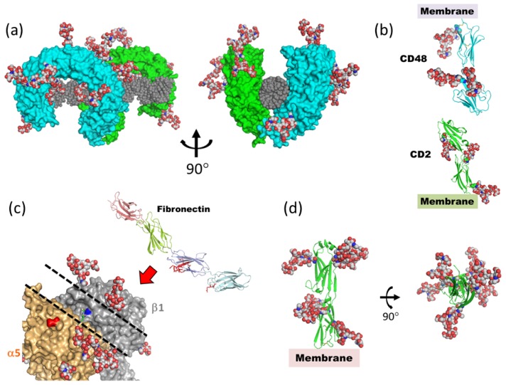
Figure 10.
Highly flexible N-glycans on cell surface receptors. Complex-type N-glycans (GlcNAc2Man3GlcNAc2Fuc) are superimposed, based on the position of chitobiose or sequons by using LSQKAB [103]. (a) Fully glycosylated Toll-like receptor-3 (TLR3) ectodomain in complex with dsRNA (PDB code; 3ciy). Protein molecules are shown as green and cyan surface models. dsRNA is shown as a gray sphere. (b) Extracellular domains of CD2 (PDB code; 1hnf) and CD48 (PDB code; 2dru). (c) Crystal structure of α5β1 integrin ectodomain (PDB code; 3vi4) and fibronectin FN7-10 fragment (PDB code; 1fnf). In the fibronectin structure, the amino acid residues which interact with α5β1 integrin are shown in red stick model. Dashed lines on α5β1 integrin outline the shallow groove formed by N-glycans. (d) Intercellular cell adhesion molecule (ICAM)-2 ectodomains (PDB code; 1zxq).

