Figure 6.
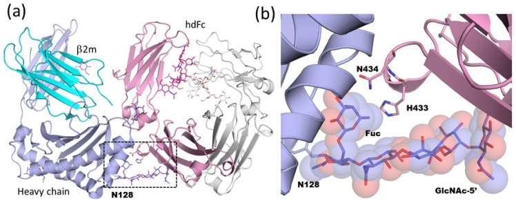
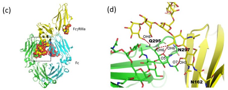
(a) Overall structure of neonatal Fc receptor (FcRn) in complex with heterodimeric Fc (hdFc) (PDB code; 1i1a). Heavy chain and soluble light chain β2-microglobulin (β2m) of FcRn are shown in slate and cyan, respectively. Proximal and distal Fc fragments of hdFc are shown in pink and white, respectively. The region delineated in black dotted lines is magnified in (b). (b) Close-up view of FcRn-hdFc complex. N-glycan attached at Asn128 of FcRn is shown in rod and semitransparent sphere model. (c) Overall structure of human Fc-glycosylated human Fcγ receptor IIIa (FcγRIIIa) complex (PDB code; 3sgk). Two chains of Fc fragment and FcγRIIIa are shown in green, cyan, and yellow, respectively. The region delineated in black dotted lines is magnified in (d). (d) Close-up view of carbohydrate-carbohydrate interaction in Fc-FcγRIIIa. Hydrogen bonds are shown as red dotted lines.


