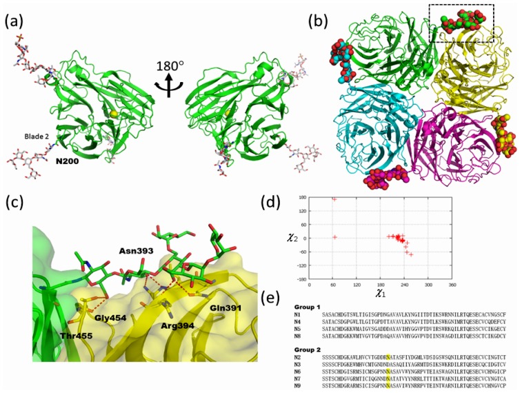
Figure 7.
High-mannose type glycan of influenza neuraminidase assists tetramer formation. (a) Overall structure of monomeric influenza N2 neuraminidase (PDB code; 1nn2). Protein, carbohydrate, and calcium ion are shown in ribbon, stick, and sphere models, respectively. (b) Tetrameric structure of influenza N2 neuraminidase (PDB code; 1nn2). N-linked glycans at Asn200 are shown in sphere models. The region delineated in black dotted lines is magnified in (c). (c) Close-up view of N-glycan at Asn200 and symmetry related molecule. Hydrogen bonds are shown in red dotted lines. (d) The side-chain torsion angles of Asn200 of N2, Asn207 of N6, and Asn200 of N9 NA (Asn201 in PDB code; 2b8h). (e) Amino acid sequence alignment of group 1 and 2 influenza neuraminidase around Asn200 glycosylation sites. Putative N-linked glycosylation sites in group 2 are highlighted.

