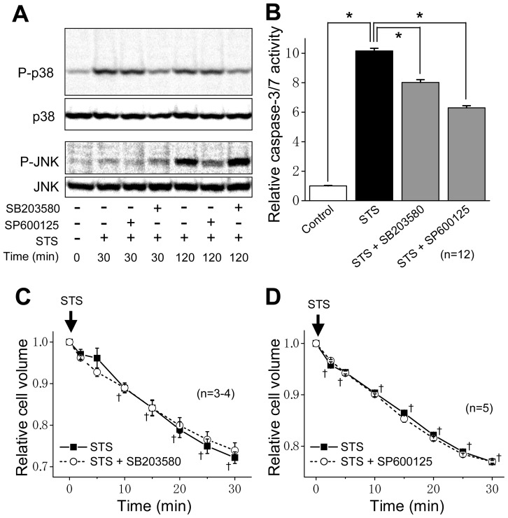
Figure 4.
Effects of a MAPK inhibitor on STS-induced MAPK phosphorylation, caspase activity and AVD in HeLa cells. (A) Western blot analysis for the levels of P-p38, p38, P-JNK and JNK in the absence (−) or presence (+) of 10 μM SB203580 or 15 μM SP600125 30 min or 120 min after STS (4 μM) stimulation. (B) Caspase-3/7 activity before (Control: open column) or after 4-h stimulation with 4 μM STS in the absence (filled column) or presence of 10 μM SB203580 or 15 μM SP600125 (shadowed columns). Each column represents the relative mean value (normalized by the control value) with SEM (vertical bar) (n = 12). * p < 0.05 between two data designated. (C, D) Time course of changes in the mean cell volume after stimulation with 4 μM STS in the absence (filled squares) or presence (open circles) of 10 μM SB203580 (C) or 15 μM SP600125 (D). Each symbol represents the relative mean cell volume at a given time (normalized by the mean cell volume at time zero), and each vertical bar represents the SEM value (n = 3–5). † p < 0.05 between the data in the absence of a MAPK inhibitor at time zero and at a given time.

