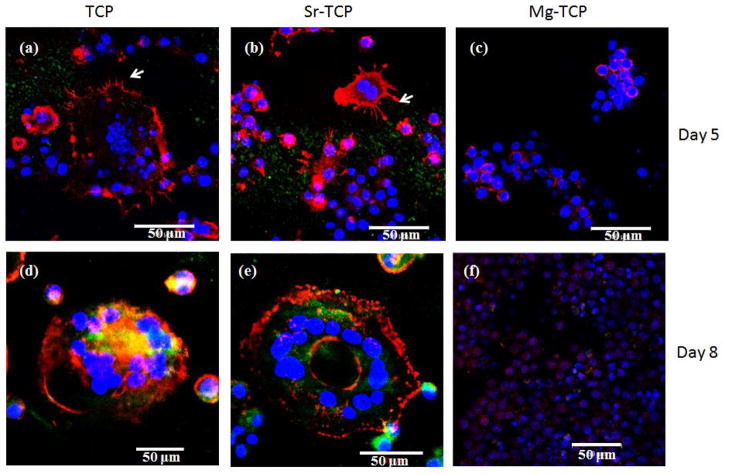
Figure 5.
Fluorescence microscopy images of cells cultured for 5 and 8 days on TCP, Sr-TCP and Mg-TCP samples. The red represents the actin cytoskeleton and the blue represents the nucleolus. The podosomes are represented by white arrow. Cells with multiple nucleuses and acting ring were identified as osteoclast-like cells.

