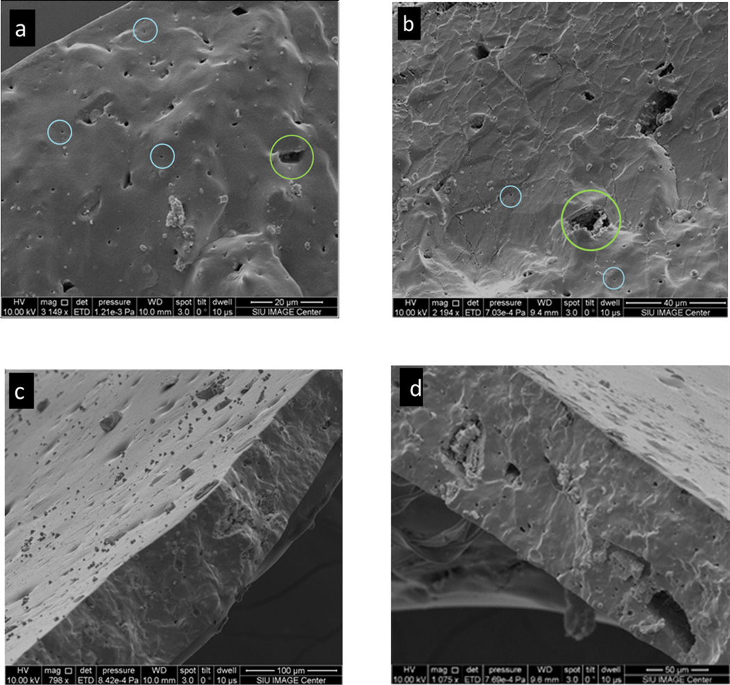
Figure 3.
Secondary electron images of the pore structure after filling the pore channels with gold nanoparticles. (a) and (b)A secondary electron image showing pores on the cross-section of the PDMS membrane. Blue and green denote the large pore and small pores on the cross-section; (c) and (d) a secondary electron image showing the pores on both the cross-section and the surface of the regular PDMS membrane.

