Figure 3 .
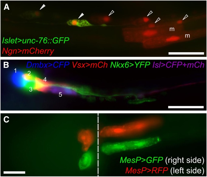
Electroporation of reporter gene plasmids. (A) Posterior region of the tail of a midtailbud-stage embryo, coelectroporated with Islet > unc-76::GFP (green) and Ngn > mCherry (red) reporter constructs. Ngn is expressed in A- and b- neural plate border cells, which gives rise to ependymal cells (open arrowheads) and bipolar neurons (solid arrowheads) of the posterior tail nerve cord, as well as secondary tail muscle cells (“m”). The Islet driver used is Islet -5915/-5396 (Stolfi et al. 2010), which only drives expression in bipolar tail neurons. This exemplifies the use of lineage- and cell-type–specific reporter plasmids to visualize cells and trace their ontogeny. Ngn > mCherry was constructed using 5 kb upstream of the endogenous Ngn start codon. (B) Motor ganglion of a C. intestinalis larva transfected with five different plasmids for a combination of four different drivers and three different fluorescent reporter genes. This mix allows for labeling of all five neuronal subtypes of the motor ganglion (numbered 1–5) in a single individual. All reporters were fused to unc-76 tag for efficient labeling of axons. mCh, mCherry; Isl, Islet. Panel adapted from Stolfi and Levine (2011). (C) Ventral view of a midtailbud-stage embryo electroporated once with MesP > GFP (green), rinsed, and electroporated with MesP > RFP (red). Due to mosaicism, this individual is expressing MesP > GFP only in cells of the right hemisphere, and MesP > RFP only in cells of the left hemisphere. Image is a composite of images taken at different focal planes of the same embryo. The seam between the two focal planes is indicated by the dashed line. Migratory heart precursors are labeled in the left panel, while tail muscles are labeled in the right panel. Both are descended from the B7.5 pair of blastomeres, which express MesP. Bars in A and B, ∼25 µm.

