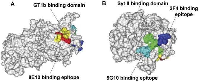
Figure 4. Molecular model overlay of neutralizing epitopes within the BoNT/B Hc binding domain.
The model was established using the software Discovery Studio 2.0 (Accelrys, San Diego, CA) based on the crystal structure of BoNT/B Hc (PDB 1F31) from the Protein Data Bank. BoNT/B Hc is shown in a surface representation. (A) The residues reported as GT1b-binding site are colored yellow (Nat. Struct. Biol. 1998), and the residues recognized by 8E10 are colored red. (B) The SytII-binding site residues are colored green, and the residues of 5G10 and 2F4 are indicated in cyan and blue.

