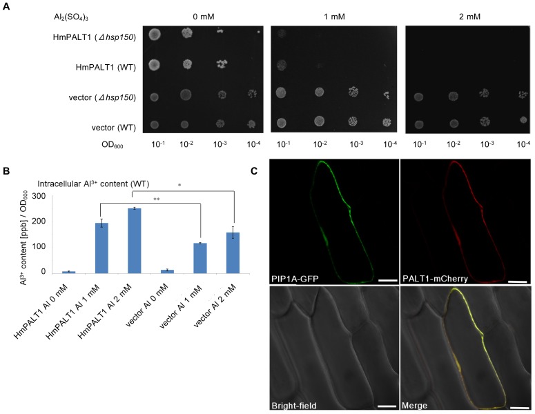
Figure 6. Identification of a plasma membrane-localized Al-transporter, HmPALT1, in hydrangea sepals.
(A) Al tolerance assay of HmPALT1. Δhsp150 and wild-type (WT) yeast cells carrying HmPALT1 or the empty vector (pYES2) were spotted onto LPP–uracil medium (pH 3.5) with or without 1 and 2 mM Al2(SO4)3, and the plates were incubated at 30°C for 4 d. (B) The intracellular Al content of yeast cells that were transformed with HmPALT1. Yeast cells carrying HmPALT1 or the empty vector were exposed to different concentrations of Al2(SO4)3 at pH 3.5 for 2 d. The intracellular Al content in both transformants increased with the Al in the medium in a dose-dependent manner. It is noteworthy, however, that the Al content of the yeast carrying HmPALT1 was approximately twice as much as that of the vector control cells, which was significant (**P<0.01 and *P<0.05 by Student's t test, respectively). The error bars represent SE (n = 3). (C) Subcellular localization of HmPALT1. The construct pHmPALT1-mCherry was simultaneously introduced into Welsh onion epidermal cells by particle bombardment with pPIP1A-GFP as a plasma membrane marker. The fluorescent signals were observed under microscopy 21 h after the bombardment. The merged image shows that HmPALT1 localizes to the plasma membrane. Scale bar = 50 µm.

