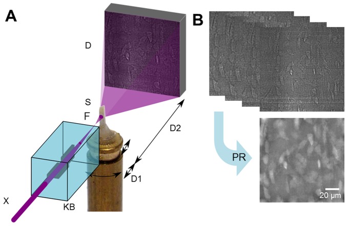
Figure 1. Experimental setup and image reconstruction.
[a] Schematic of experimental setup. The X-ray beam [X] is monochromatized and focused into a focal spot [F] by X-ray reflective optics [KB]. The sample [S] is positioned on a translation-rotation stage downstream of the focus and imaged onto a stationary detector. Due to the resulting divergent beam, different spot-sample distances [D1] and different free space propagation distances [D2] imply different magnification factors on the detector. [b] Images were recorded at four focus-to-sample distances over a complete turn of the sample at 2999 projection angles. The images were used to reconstruct the phase shift at each angle [phase retrieval PR], which was used as input to a tomographic reconstruction algorithm to reconstruct the 3D local mass density.

