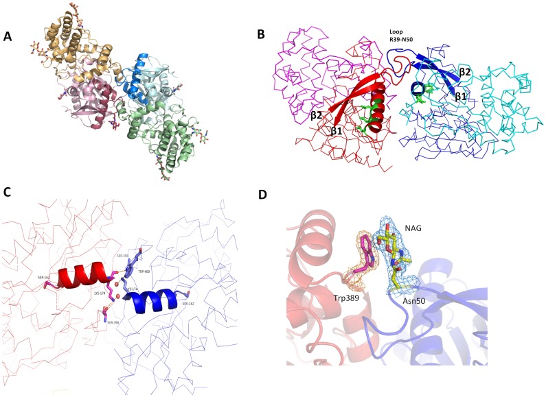
Figure 2. The DPP7 dimerization interface.
(A) Cartoon representation of DPP7 overall structure highlighting the two protomers: helical domains are shown in orange and green, the α/β-hydrolase domains in red and blue. Carbohydrates are shown as sticks. (B) Dimerization mediated by the loop Arg39 – Asn50. Ribbon representation with two strands of the central β-sheet and the loop Arg39 – Asn50 represented as cartoon. The protomers are colored in blue and red, respectively. The supposed leucine zipper motifs are highlighted as cartoon and shown in green. (C) Dimerization mediated by helix α5. The catalytic Ser162 and residues participating in hydrogen bonds at the other of the helix are represented as sticks. Two water molecules are shown as red spheres. Helix α5 is represented as cartoon and the protomers are represented as ribbons in red and blue. (D) Stacking interaction between N-acetylglycosamine (NAG) linked to Asn50 and Trp389. A 2Fo-Fc electron density map of NAG and Trp389 is shown contoured at 1 σ. The figure was prepared using the program PyMOL (http://www.pymol.org/).

