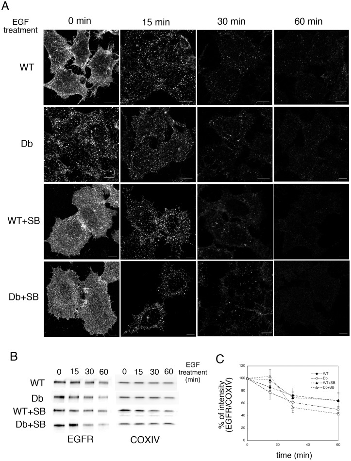
Figure 7. Degradation of EGFR in WT and Db cells.
A. HeLa cells were preincubated with DMEM without serum overnight, and were incubated without (WT and Db) or with (WT+SB and Db+SB) 2 µM SB203580 for 60 min. Semi-intact HeLa cells were incubated with WT or Db liver cytosol that contained Alexa546-conjugated dextran, and were resealed. After incubation with DMEM in the presence or absence of SB203580 for 30 min, the cells were treated with 10 ng/ml EGF, and then incubated with medium at 37°C for 0, 15, 30, and 60 min. The cells were fixed, and stained with anti-EGFR antibody. We were able to distinguish resealed cells, which were fluorescently labeled with dextran, from non-resealed cells easily under a fluorescence microscope. Bar = 10 µm. B. HeLa cells were treated as described in A, lysed, and subjected to Western blotting using antibodies against EGFR and COX VI. C. Means and standard deviations for the band intensities of EGFR/COX VI are shown in the graph. We performed three independent experiments and verified the results by applying Student’s t-test. We found that the P value was > 0.05, which indicated that treatment with 2 µM SB203580 did not affect the enhanced degradation of EGFR by Db cytosol.

