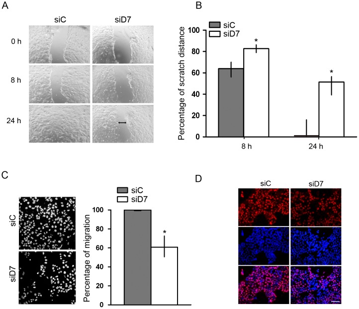
Figure 3. Effect of StarD7 silencing on JEG-3 cell migration.
A- Wound healing assay in JEG-3 cells treated with StarD7.1 (siD7) or scrambled (siC) siRNAs. An open furrow was generated by scratching confluent cells using a pipette tip. Confluency was restored in controls after 24 h. However, in cells treated with StarD7 siRNA, confluency was not restored after 24 h. B- The distance between furrow edges in the scrambled or StarD7 siRNA-treated cells of three independent experiments was measured and presented graphically as percentage of the initial distance (0 h);*p<0.05 compared to scrambled siRNA-transfected cells. C- Transwell In vitro migration assays. Left panels: representative images show cells migrated to the lower chamber after 48 hours (×200). Right panels: Bar graph represents the percentage of cell migration in seven fields of duplicate wells containing StarD7 siRNA-treated cells relative to scrambled siRNA ones (median and 25th–75th% percentiles, n = 3); *p<0.05 compared to scrambled siRNA-transfected cells. D-Cell proliferation was determined by BrdU (red) staining of JEG-3 cells treated with scrambled (left panel) or StarD7.1 (right panel) siRNAs. The nuclei were labelled with Hoescht (blue, middle panels) and merge images are shown on the bottom panels. Bar = 50 µm (×200). The images are representative of three experiments with consistent results.

