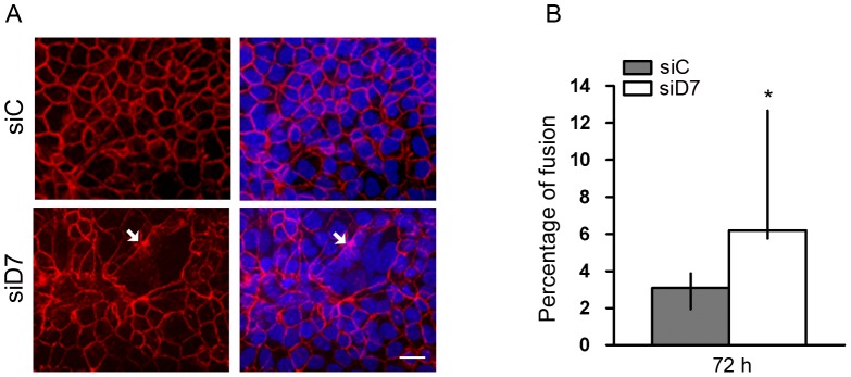
Figure 7. Effect of StarD7 knockdown on JEG-3 cell differentiation.
Cells were transfected with scrambled (siC) or StarD7.1 (siD7) siRNAs for 6 h and then cultured until 72 hours. A- Detection of desmoplakin (red) in siRNA-treated JEG-3 cells was performed by immunofluorescence (left panel). The nuclei were labelled with Hoescht (blue) and merge images are shown on the right. Syncytial structures were characterized by the absence or incomplete desmoplakin staining (arrows). In control cells, desmoplakin labeling was continuous at the periphery of cells. Bar = 20 µm (×400). B- Percentage fusion was determined in cells transfected with StarD7.1 or scrambled siRNAs and represents the percentage of nuclei number in syncytia. Twenty fields were counted for each condition in three different experiments performed in duplicate as described in Materials and Methods. Results are depicted in terms of median percentage and 25th–75th% percentiles; *p<0.05 compared to scrambled siRNA-transfected cells.

