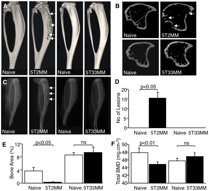
Figure 1. 5T2MM, but not 5T33MM cells cause osteolytic bone disease and tumour-induced bone loss.
A. Reconstructed 3-dimensional micro-CT images of the tibia of naïve mice, mice bearing 5T2MM cells and mice bearing 5T33MM cells. Lesions in the tibia of 5T2MM bearing animals are arrowed. B. Transverse sections of micro-CT images of tibiae from naïve mice, mice bearing 5T2MM cells and mice bearing 5T33MM cells. Lesions are arrowed. C. Radiographs of the tibia of naïve mice, mice bearing 5T2MM cells and mice bearing 5T33MM cells. Lesions are arrowed. D. Number of lesions in the tibia of naïve mice and 5T2MM or 5T33MM bearing mice. E. Trabecular bone area as a proportion of total tissue area in the tibia of naïve mice, and mice bearing 5T2MM or 5T33MM cells. F. Total bone mineral density of naïve mice, and 5T2MM or 5T33MM bearing mice. Statistical analysis by Mann-Whitney U test. Data = mean± S.E.M.

