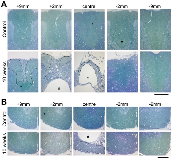
Figure 1. Plastic sections (0.5 µm) stained with methylene blue of aged matched control and at 10 weeks after injury rat spinal cords.
(A) dorsal column, (B) ventrolateral tracts at different distances from injury centre. The #-symbol marks the cyst that develops after the injury. In the dorsal column at 10 weeks after injury only a small area is left in which axons are visible (area marked with dotted line); this area becomes progressively larger rostrally. Caudal to the injury the dorsal column is little affected; however, the corticospinal tract that runs at the base of the dorsal column (marked with an asterix) is not visible caudal to the injury but in contrast appears normal rostrally. Scale bars are 300 µm.

