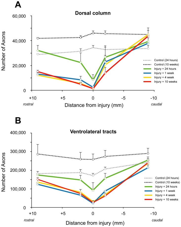
Figure 5. Number of myelinated axons in the dorsal column (A) and ventrolateral tracts (B) in control and spinal injured rats.
A) In the middle of the injury site at 24 hours after the injury, the number of axons is reduced to 23% of controls (0 week) and further reduced to 4–6% at later times after injury. The number of axons was similar in animals at 1–10 weeks after the injury at all distances from injury and was much lower than controls even at 9 mm rostral to injury (43–52% of 0 week controls) whereas at 9 mm caudal numbers were similar to controls. Note that in 10-week controls the number of axons is about 38% (or 12,000 axons) more than in 0-week controls in the dorsal column. B) In the middle of the injury site at 24 hours after the injury, the number of axons is reduced to 54% of controls (0 week) and further reduced to 13–17% at later times after injury. The number of axons was similar in animals at 1–10 weeks after the injury at all distances from injury and was lower than controls at 9 mm rostral (71–83% of 0 week controls) whereas at 9 mm caudal it was similar to controls. Note that in 10 week controls the number of myelinated axons is about 40% (or 76,000 axons) more than in 0 week controls in ventrolateral tracts.

