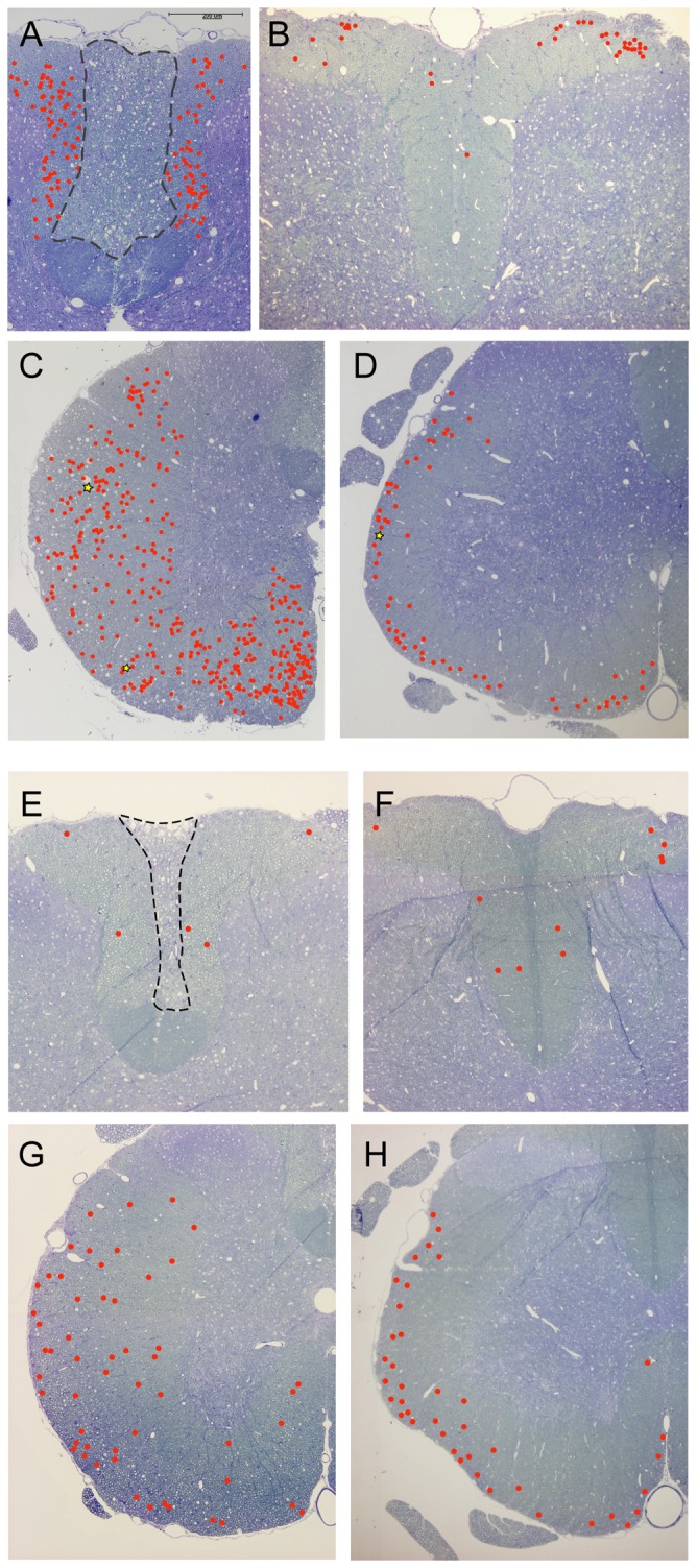
Figure 8. Illustrations of the general distribution of mapped axonal pathology at 1 week (A–D) and 10 weeks (E–H) after injury.

Axons with moderate pathology are shown with red circles and axons with severe pathology with yellow stars. For illustrations of pathology see Figure 7. A) Axons in the dorsal column, 9 mm rostral of injury. Note that the pathology is uniformly spread throughout the white matter, outside the central core that is filled with degenerated axons (area inside dashed line). B) Axons in the dorsal column, 9 mm caudal of injury. Axon pathology is visible almost exclusively in the dorsolateral parts of the column where the axons enter from the spinal roots. C) Axons in the ventrolateral tracts, 9 mm rostral of injury. Axons pathology is uniformly spread within the white matter with some concentration to the medial parts. D) Axons in the ventrolateral tracts, 9 mm caudal of injury. Axon pathology is mostly restricted to the outer layers of the white matter close to the pial rim. Note that the numbers are much less than rostral to the injury. E) Axons in the dorsal column, 9 mm rostral of injury. A few axons are found outside the central core that is filled with degenerated axons (area inside dashed line). F) Axons in the dorsal column, 9 mm caudal of injury. A few axons are found throughout the column. G) Axons in the ventrolateral tracts, 9 mm rostral of injury. Axons pathology is rather uniform within the white matter. H) Axons in the ventrolateral tracts, 9 mm caudal of injury. Axon pathology is concentrated towards the pial rim in white matter and numbers are similar to caudal to injury (G).
