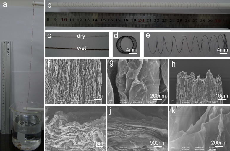
Figure 1. Preparation and microstructures of macroscopic GO fibers.
Photographs of (a) 50 cm-long fiber drawn out from the CTAB solution by wet spinning of the 10 mg/mL GO dopes in a coagulation bath of 0.5 mg/mL CTAB solution; (b) ~ 1.6 m-long fiber wound on a Teflon rod with a diameter of 8 mm; (c) the dry and wet fibers; (d) the fiber coil; (e) spring-like fiber by spreading out of coil in (d). (f, g) SEM images of the axial outer surface with different magnifications. (h–k) SEM images of the end of the broken part the fiber with different magnifications.

