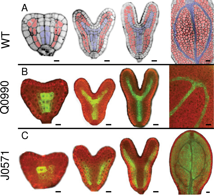
Fig. 1.
GFP expression in vascular (Q0990) and ground (J0571) tissues during embryo development. Confocal images show vascular tissues (blue) and ground tissues (red) in wild-type embryos (A) or GFP expression in, Q0990 (B) and J0571 (C) embryos at different developmental stages (left to right: triangular/early heart, late heart, torpedo, mature embryonic cotyledon). All images are median longitudinal sections except the early heart stage of J0571, which is a tangential longitudinal section taken through the ground tissue showing GFP expression in these cells. Bars, 10 µm.

