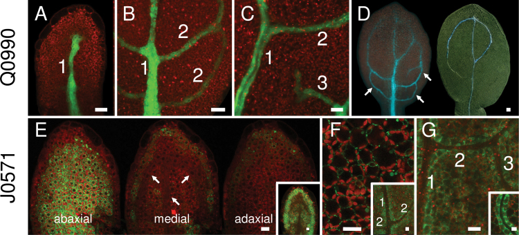
Fig. 2.
GFP expression in vascular (Q0990) or ground (J0571) tissues during leaf development. Images show first rosette leaf primordia at 2 (A), 3 (E), 4 (B, F inset), 5 (C, D, G), 6 (F), and 7 (G inset) days after germination. (A–C) GFP expression in Q0990 leaf primordia was observed progressively in the primary midvein (1), secondary veins (2), and tertiary veins (3). (D) In Q0990, GFP fluorescence (left) and cleared darkfield image of the same leaf showing differentiated xylem (right) indicate that GFP expression was also present in undifferentiated procambial cells (arrows). (E–G) In J0571, GFP expression was initially mostly in abaxial tissue (arrows in medial section show procambial cells) (E) and progressively included more adaxial ground tissues (E inset, F), increasingly delimiting venation (F inset, G), and eventually becoming more prominent in cells directly surrounding the vascular tissue (G inset). Bars, 10 μm (A–C, E–G), 20 µm (D).

