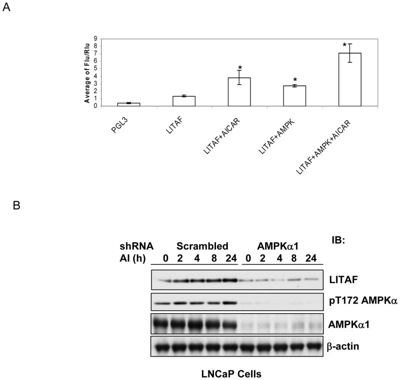
Figure 1. AMPK-dependent induction of LITAF by AICAR.
A. Promoter activity assay. LNCaP cells were transfected with a promoter-less fire fly luciferase reporter plasmid (PGL3) or the plasmid with insertion of LITAF promoter region and Renilla luciferase plasmid in the presence or absence of a plasmid encoding wild type AMPK α1 subunit. Two days later, the cells were treated with AICAR for 8 hours and luciferase assay was conducted as described in Materials and Methods. The luciferase activity was normalized with Renilla lucicerase activity and expressed as ratios (Means ± SD, n=3). “*” denotes P<0.01 as compared to basal LITAF promoter activity. B. LNCaP cells stably infected with α1 shRNA or scrambled shRNA retrovirus were treated with AICAR (AI, 1 mM) for indicated times. Cell lysates were blotted with antibodies, as indicated.

