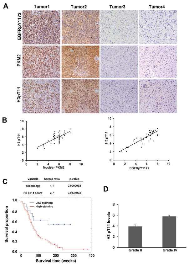
Figure 6. H3-T11 Phosphorylation Positively Correlates with the Level of Nuclear PKM2 Expression and with Grades of Glioma Malignancy and Prognosis.

(A, B) Immunohistochemical staining with anti–phospho-EGFR Y1172, anti–phospho-H3-T11, and anti-PKM2 antibodies was performed on 45 GBM specimens. Representative photos of four tumors are shown (A). Semiquantitative scoring was performed (Pearson product moment correlation test; r = 0.704, p < 0.0001, left panel; r = 0.86, p < 0.001, right panel). Note that some of the dots on the graphs represent more than one specimen (some scores overlapped) (B).
(C) The survival times for 85 patients with low (0-4 staining scores, blue curve) versus high (4.1-8 staining scores, red curve) H3-T11 phosphorylation (low, 16 patients; high, 69 patients) were compared. The table (top panel) shows the multivariate analysis after adjustment for patient age, indicating the significance level of the association of H3-T11 phosphorylation (p = 0.01349038) with patient survival. Empty circles represent deceased patients, and filled circles represent censored (alive at last clinical follow-up) patients.
(D) Thirty diffuse astrocytoma specimens were immunohistochemically stained with anti–phospho-H3-T11 antibody, and specimens were compared with 45 stained GBM specimens (Student’s t test, two tailed, p < 0.001). Data represent the mean ± SD of and 30 stained astrocytoma specimens and 45 stained GBM specimens.
