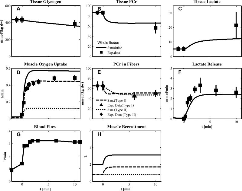
Figure 6.
Simulated dynamics and experimental data for intracellular species concentrations in muscle in response to an increased work rate equivalent to one-leg knee-extensor exercise at 50% VO2max for subjects corresponding to Experiment 4: (A) Glycogen; (B) Phosphocreatine (PCr); (C) Lactate in tissue; (D) Muscle oxygen uptake; (E) PCr in type I and II fibers; (F) Lactate release; (G) Blood flow (Q); (H) Effective volume change of whole muscle, type I and II fibers and blood domain. The blood flow curve was obtained by data interpolation. Lines represent model simulations of whole muscle (—), type I fibers (- - -), and type II fibers (. . .). Experimental data are means ± standard error. Solid squares for whole tissue; upper triangles for type I fibers; diamonds for type II fibers.

