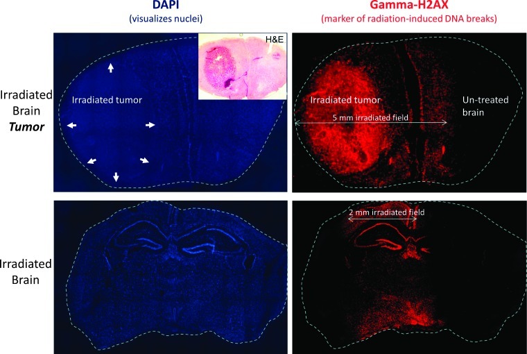
Figure 3.
γH2AX staining of irradiated brain tissue confirms accurate delivery of stereotactic brain irradiation. Top row shows fluorescent microscopy images of coronal brain sections from a mouse with an intracranial (forebrain) tumor (marked with arrows) killed 2 hours after focal RT to 20 Gy in a single fraction using a 5 x 5-mm collimator, followed by staining for DAPI to visualize cell nuclei (left) and γH2AX to visualize radiation-induced unrepaired double-strand breaks (right). γH2AX staining confirms the precise targeting of the tumor using the described technique. The bottom row shows DAPI and γH2AX staining performed on coronal midbrain sections of a mouse without intracranial tumor killed 2 hours after focal RT to 20 Gy x 1 using a 7 x 2-mm collimator with the long axis parallel to the animal's spine.

