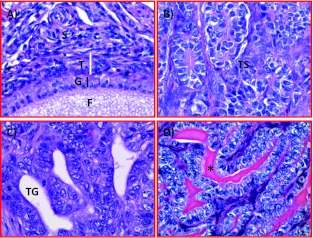
Figure 2.
Microscopic features of hen ovaries. Paraffin-embedded sections from normal or malignant ovaries with tumors were stained with hematoxylin and eosin. (A) Section of a normal ovary showing a developing follicle embedded in the ovarian stroma. (B) Section of an ovarian serous carcinoma showing a solid sheet of tumor surrounded by fibromuscular tissue. The tumor contains a labyrinth of slitlike glandular spaces lined by cells with large pleomorphic nuclei and mitotic figures. (C) Section showing endometrioid carcinoma displaying confluent back-to-back glands. Glands contain a single layer of epithelial cells with sharp luminal margins. (D) Section of a mucinous carcinoma. Glands in clusters with scarce intervening stroma lined by columnar and goblet cells with intracytoplasmic mucin. Original magnification, x40.

