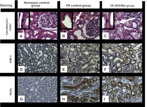
Fig. 3.
Effect of αKG/MAL on kidney histopathology (hematoxylin-eosin staining, A–C), immunohistochemical staining of kidney injury molecule-1 (KIM-1; D–F), and neutrophil gelatinase-associated lipocalin (NGAL; G–I) following kidney I/R. Anesthetized rats were subjected to bilateral kidney ischemia (I; 40 min) and subsequent reperfusion (R; 240 min). αKG/MAL was infused at 0.0095 mmol·kg−1·min−1 each. Normoxic and I/R control group animals received only 0.9% NaCl solution (5 ml·kg−1·h−1). Representative pictures at 400-fold magnification are shown. A, D, and G: normoxic control group. B, E, and H: I/R control group. C, F, and I: I/R αKG/MAL group. Stainings were performed at the end of the experimental procedure. Arrows in B and C indicate vacuolization. Insets: damage to brush border.

