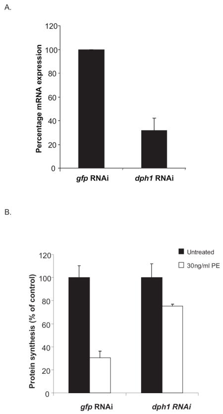
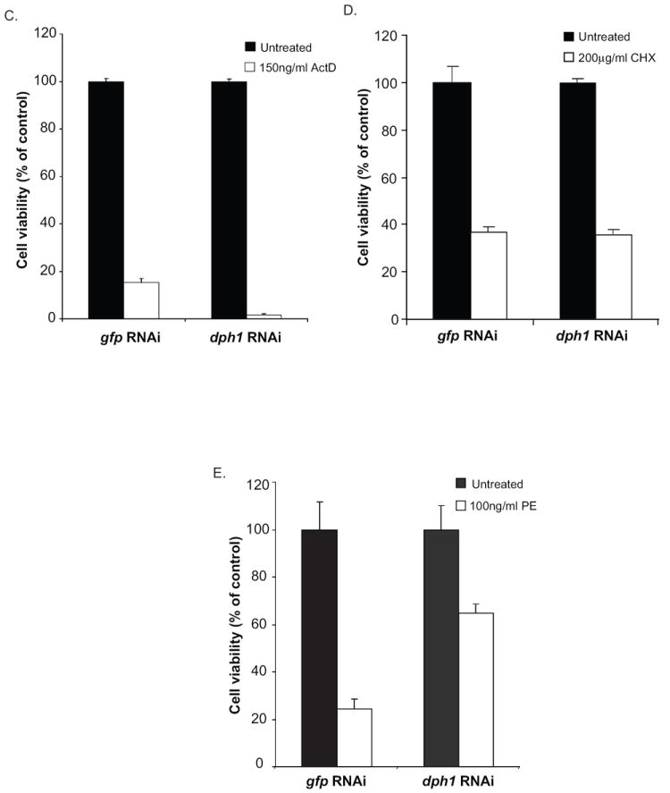
Fig. 2.


dph1 knockdown prevents both PE-mediated inhibition of protein synthesis and cell death. (A) S2 cells were incubated with dsRNA to dph1 or a non-target gene, gfp, and the expression of dph1 mRNA measured using real time PCR. Rpl32 expression was used as an internal control. The data are expressed as means of three independent experiments. (B) S2 cells were subjected to RNAi to dph1 or a control gene, followed by an incubation with 30ng/ml PE for 24 h. Protein synthesis inhibition was assessed by measuring the amount of {3H}-leucine incorporated into cellular material and is presented as the percentage value of an untreated sample. S2 cells were subjected to RNAi using dsRNA to dph1 followed by an incubation with 150ng/ml actinomycin D (C), 200μg/ml cycloheximide (D) or 100ng/ml PE (E) for 48 h. Cell viability was measured using alamarBlue® and the data is presented as percentage value of the untreated sample. Each bar is the mean of at least three determinations. Error bars represent one standard deviation from the mean.
