
Fig. 3.
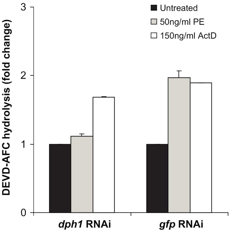
PE treatment leads to activation of caspase activity. S2 cells were subjected to RNAi using dsRNA to dph1 followed by incubation in the presence of 50ng/ml PE or 150ng/ml actinomycin D for 24 h. dsRNA to a non-specific target gene, gfp, was used as control. An increase in the caspase (presumed to be Drice, see below) activity was estimated by measuring the increase in fluorescence upon hydrolysis of DEVD-AFC. The specific caspase activity of the dph1 knockdown samples is presented as the fold-change value compared to an untreated sample. Each bar is the mean of at least three determinations. Error bars represent one standard deviation from the mean.