Figure 1.
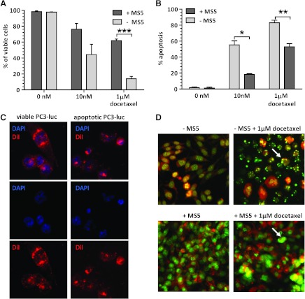
(A) Viability of PC3-luc cells after docetaxel treatment with or without the presence ofMS5 stromal cell line. (B) Apoptosis of PC3-luc cells after docetaxel treatment with or without MS5 stromal cell line. (C) Exemplary figures presenting the assessment of PC3-luc cell viability by fluorescence microscopy (40x). The nuclei (blue, DAPI) are round and intact with visible nucleoli in viable cells, whereas nuclei of apoptotic PC3-luc cells are condensed and fragmented. Cytoplasm visualized in red (DiI) shows regular structure in viable cells, whereas it is condensed in apoptotic cells. (D) Exemplary pictures representing the assessment of apoptosis of PC3-luc cell line obtained by fluorescence microscopy (40x magnification). Apoptotic cells, defined by chromatin condensation and presence of apoptotic bodies, are indicated by arrows. *P < .05, **P < .005, ***P < .0005.

