Figure 3.
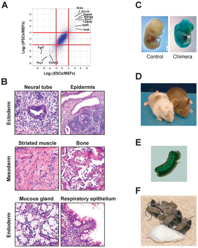
Verification of pluripotency of mouse M3O-induced pluripotent stem cells (iPSCs). (A): Expression level of transcripts in M3O-iPSCs and embryonic stem cells (ESCs) relative to mouse embryonic fibroblasts (MEFs). Log2 ratios are plotted for transcripts in ESCs/MEFs and iPSCs/MEFs. Red lines indicate a fourfold difference in transcript levels. Transcripts in M3O-iPSCs were assayed 60 days after transduction. (B): H&E staining of teratoma sections derived from M3O-iPSCs. Neural tube and epidermis (ectoderm), striated muscle and bone (mesoderm), and mucous gland and respiratory epithelium (endoderm) are shown. Scale bar = 50 μm. (C): X-gal staining for cells expressing the lacZ gene in a chimeric embryo prepared with M3O-iPSCs and a control embryo at 13.5 dpc. (D): Chimeric mice prepared with M3O-iPSCs. The agouti coat color indicates a high (right) and low (left) contribution of iPSCs to the skin. The host embryos used to generate mice were derived from the albino mouse strain ICR. (E): Germline contribution of M3O-iPSCs as shown by green fluorescence protein expression in the gonad of a 13.5-dpc chimeric embryo. (F): Pups obtained from crossing a wild-type ICR female (bottom) with an M3O-iPSC chimeric male (left mouse in panel D). Abbreviations: ESCs, embryonic stem cells; iPSCs, induced pluripotent stem cells; MEFs, mouse embryonic fibroblasts.

