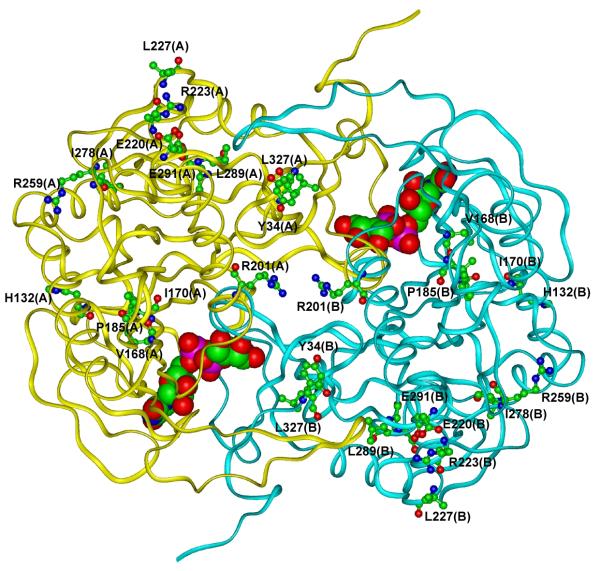
Fig. 3. Schematic view of the modeled 3D structure of wild-type GALT enzyme.
The backbone of the protein is shown as a ribbon. UDP-Gal is shown in CPK mode and residues that undergo the variations are represented in ball & stick mode and labeled. Hydrogen atoms are not represented for sake of clarity. (In the color version online, the two subunits forming the dimer are colored in yellow and cyan for monomer A and B, respectively. Color codes for atom types are: carbon: green, oxygen: red, nitrogen: blue, phosphorus: magenta).

