Figure 5.

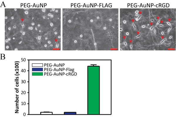
Enhanced adhesion of HeLa cells to the cRGD-doped nanofiber substratum. AuNPs labeled with cRGD and PEG were used to dope the PMGI nanofibers through electrospinning, with which a synthetic substratum was fabricated. The same number of HeLa cells were seeded and incubated for 6 h over the cRGD-doped nanofiber substrata and other nanofiber substrata containing plain AuNPs or FLAG-labeled AuNPs. The red arrows indicate attached and spread HeLa cells (a). After washing the cells with PBS, the remaining cells were collected from the 3 dishes of each substratum via trypsinization for quantification (b). Scale bar: 50 μm. Error bars: SEM, n = 3.
